Research materials
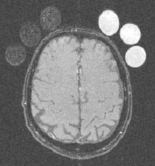
Brain cross-sectionTo compare properties of different phantoms, in GCRC, Heidelberg a series of MRI images were measured. They contain cross-sections of 3 polistyren phantoms with different bead diameters, 3 different porosity foam phantoms and cross-section of human brain. Example of this image is shown in Fig. 6.
Currently, a research is going on to find a feature set able to classify the analysed class of MRI textures, representing different phantoms and human brain tissues. For this purpose, a specialised software package was developed under B11 COST action. << Back | Kidney image >> |